

HUMAN BRAIN
HUMAN BRAIN:
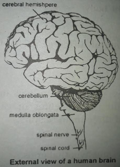
Brain is the most complex and advanced part of the human body. It is the coordinating Centre present in the cranium of the skull.
LOCATION:
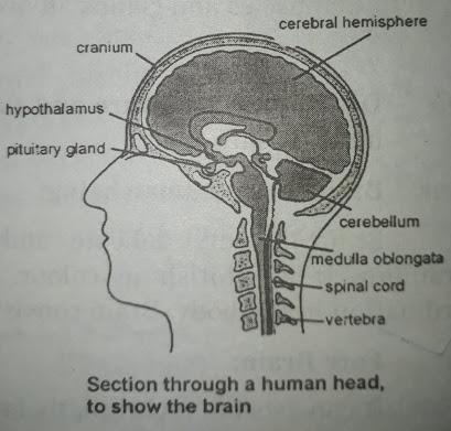
It lies in a cavity called cranium supported by a particular called meninges.
CRANIAL NERVES:
Nervous arising from the brain are called cranial nerves. There are 12 pairs of cranial nerves arising from the brain.
PARTS OF BRAIN:
Human brain can be divided into three parts.
1) Fore brain
2) Midbrain
3) Hindbrain
1. FORE BRAIN:
The first part of the brain is called the brain; it is differentiated into three parts.
a)Cerebrum
b)Thalamus
c)Limbic system
a. Cerebrum:
It is the interior longest part of the brain. It is divided into two halves by a group called median fisher. Each part of the cerebrum is called the cerebral hemisphere. These hemispheres are connected with each other by Corpus callosum. The upper part of the cerebrum is called the cerebral cortex.
FUNCTION:
It receives sensory information, processes, and stores some memory for future use.
b. THALAMUS:
Thalamus is an oval shaped structure present in the arc of the cerebrum.
c. LIMBIC SYSTEM:
The Limbic system is located in an Arc between thalamus and cerebrum. It contains hypothalamus, amygdala and hippocampus. At the end of hypothalamus pituitary gland is attached.
FUNCTION:
It controls the response like hunger, thirst, fear, anger, pleasure and sexual response.
2). MIDBRAIN:
It is located between the forebrain and hindbrain. It is the smallest part of the brain.
FUNCTION:
It connects the forebrain with the hindbrain. It also controls reflex moment of eye and hearing.
3).HINDBRAIN:
It is the last part of the brain. It is divided into three parts.
a) Cerebellum
b) Pons
c) Medulla Oblongata
a). CEREBELLUM:
It is the second largest part of the brain.
FUNCTION:
It controls the balance of the body and co-ordinate the voluntary movement of the body as well.
b). PONS:
It is located above medulla oblongata.
FUNCTION:
It controls transition between sleep and wakefulness and also controls the rate of breathing.
c). MEDULLA OBLONGATA:
It is the posterior part of the brain connected with the spinal cord.
FUNCTION:
It controls involuntary function like breathing, heart rate, circulation of blood, blood pressure, swallowing and vomiting.
SPINAL CORD:

Spinal cord is a coordinating Centre present in backbone. It is the second important part of CNS.
LOCATION:
It lies in the neural Canal of the vertebral column.
SIZE:
It is 18 inches long.
SPINAL NERVES:
The nerves arising from the spinal cord are called spinal nerves. There are 32 pairs of nerves arising from the spinal cord. Spinal cord is covered by a membrane card. The fluid present between the membranes of meninges and cerebrospinal fluid.
PARTS:
Spinal cord consists of two parts.
a) Gray Matter
b) White Matter
a). Gray Matter:
The inner butterfly shaped part is called grey matter. It is grey in color.
b). White Matter:
The outer whitish part is called white matter.
FUNCTION:
● It receives messages from the body and sent to the brain.
● It receives commands from the brain and sends them to effectors.
● It controls the reflex action of the body.
REFLEX ACTION IN BODY REFLEX ACTION:
The action that is automatic, immediate, quick, involuntary and fixed response to stimulus is called reflex action.
Brain is not involved in the reflex section.
Some of reflex section are:
● blinking of eyes when something is coming to eye.
● Knee jerk when it is hit with a hard object.
● watering in mouth by seeing some delicious food.
● Retracting of hand from hot object.
REFLEX ARC:
The pathway of reflex action is called reflex arc. This action is performed by the connection of three neurons making an arc.
This starts with a sensory neuron (receptor) which takes the impulse to the spinal cord where an associate neuron shifts the impulse to the motor neuron which takes the impulse to the effector (muscle and gland).
FOR EXAMPLE:
To understand a reflex action and reflex arc. We took an example of a girl sewing a shirt with a needle. Needle is pricked into her fingers. She at once retracts hand. When the needle pricks her fingers, the receptors in the skin take this impulse to the spinal cord. There an associative neuron plugs this stimulus to a motor neuron. Motor neurons bring signals to the muscles of the hand to retract.
Other examples are blinking Of the eye, watering of mouth, knee jerk etc.


0 Comments
If you have any doubts, please let me know